When people – and monkeys – look at faces, a special part of their brain that is about the size of a blueberry “lights up.” Now, the most detailed brain-mapping study of the area yet conducted has confirmed that it isn’t limited to processing faces, as some experts have maintained, but instead serves as a general center of expertise for visual recognition.
Neuroscientists previously established that this region, which is called the fusiform face area (FFA) and is located in the temporal lobe, is responsible for a particularly effective form of visual recognition. But there has been an ongoing debate about whether this area is hard-wired to recognize faces because of their importance to us or if it is a more general mechanism that allows us to rapidly recognize objects that we work with extensively.
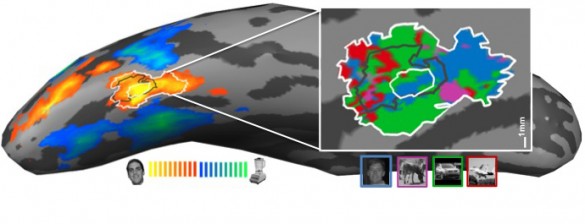
In the new study published this week in the online early edition of the Proceedings of the National Academy of Sciences, a team of Vanderbilt researchers report that they have recorded the activity in the FFAs of a group of automobile aficionados at extremely high resolution using one the most powerful MRI scanners available for human use and found no evidence that there is a special area devoted exclusively to facial recognition. Instead, they found that the FFA of the auto experts was filled with small, interspersed patches that respond strongly to photos of faces and autos both.

“We can’t say that the same groups of neurons process both facial images and objects of expertise, but we have now mapped the area in enough detail to rule out the possibility of an area exclusively devoted to facial recognition,” said Rankin McGugin, who conducted this research as part of her doctoral dissertation.
According to Isabel Gauthier, the David K. Wilson Chair of Psychology, who directed the study, the demonstration that the FFA can support expertise for other categories may help scientists improve treatments for people who have difficulty recognizing faces, such as individuals with autism. In addition, identifying the neural basis of individual differences in learning visual skills is an important step toward mapping the brain chemistry involved in learning and may eventually lead to the development of drugs that make it easier for individuals to acquire different kinds of visual expertise.
For most objects, research has shown that people use a piecemeal identification scheme that focuses on parts of the object. By contrast, experts, for faces or for cars, use a more holistic approach that is extremely fast and improves their performance in recognition tasks.
The scientists point out that visual expertise may be more the norm than the exception: “It helps the doctor reading X-rays, the judge looking at show dogs, the person learning to identify birds or to play chess; it even helped us when we learned brain anatomy!” Gauthier said.

Gauthier and her colleagues have further found that people who are better at learning to recognize one subject should also be better at learning another. Recent work by her group found that those who did a better job at identifying objects in which they were most interested were also better at identifying faces.
Christopher Gatenby at the University of Washington and John Gore, Hertha Ramsey Cress Chair in Medicine, University Professor of Radiology and Radiological Sciences and director of Vanderbilt’s Institute of Imaging Science, also contributed to the study. The research was supported by the James S. McDonnell Foundation, National Science Foundation grant SBE-0542013, National Eye Institute grant EY013441-06A2 and the Vanderbilt Vision Research Center.