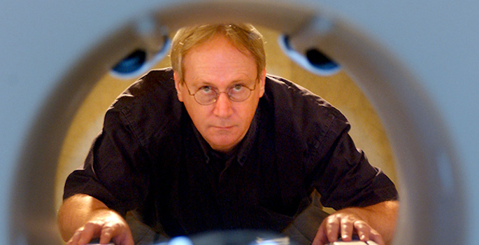
John Gore
-

Vanderbilt researchers develop comprehensive guide for how the brain’s wiring changes with age
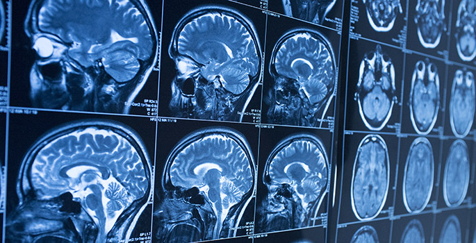
In a groundbreaking study published recently in the journal Nature, researchers at Vanderbilt University and Vanderbilt Health have created the first growth charts for white matter in the brain over a human lifetime. The work brings together nearly two decades of Vanderbilt research collaborations, the university’s extensive MRI data collections, and an advanced AI-enabled computing platform. Read MoreMay 27, 2026
-
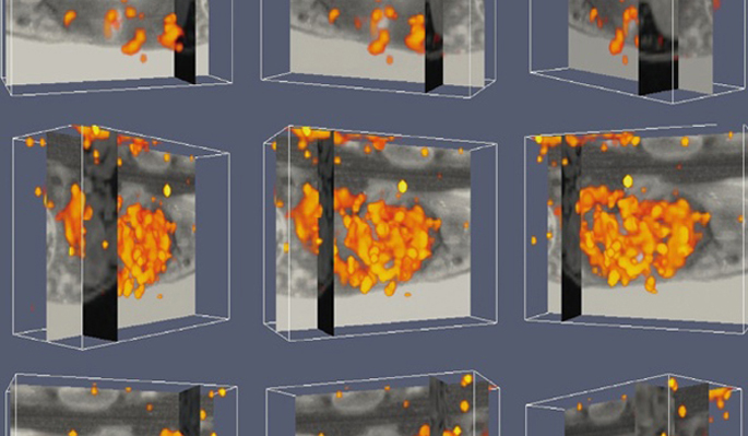
3D brain mapping opens a window to the aging brain
By mapping brain activity in three dimensions, researchers at Vanderbilt University Medical Center have achieved a more detailed picture of how the brain changes with age. Read MoreJan 30, 2024
-
VUMC scientists record powerful signal in the brain’s white matter
Vanderbilt researchers report that when people who are having their brains scanned by fMRI perform a task, like wiggling their fingers, certain signals increase in white matter throughout the brain, which has long been thought to play a lesser role the more the brain's more energetic gray matter. Read MoreOct 16, 2023
-
Imaging “biomarker” for Alzheimer’s disease progression
Changes in connectivity in the brain’s white matter may be a novel neuroimaging biomarker for assessing Alzheimer’s disease progression. Read MoreNov 16, 2020
-
Turning Heads: The Vanderbilt Brain Institute has emerged as a hub of discovery as neuroscience’s influence expands
The VBI recently marked its 20th anniversary, a span that has seen the institute’s wide-ranging missions—including administering the university’s Neuroscience Graduate Program, as well as postdoctoral training and community outreach—steadily coalesce under a single umbrella. Read MoreAug 5, 2020
-
Gore named to committee on worker health overseas
John Gore, director of the Vanderbilt University Institute of Imaging Science, has been appointed to a National Academies of Sciences, Engineering and Medicine standing committee to advise the Department of State on unexplained health effects on U.S. government employees and their families at overseas embassies. Read MoreDec 12, 2019
-

Neuromodulation device studied as non-addictive option for chronic pain
With $3.6 million in funding, researchers from the Vanderbilt University Institute of Imaging Science are developing a focused ultrasound neuromodulation device as a non-invasive and non-addictive method for treating chronic pain. Read MoreNov 11, 2019
-
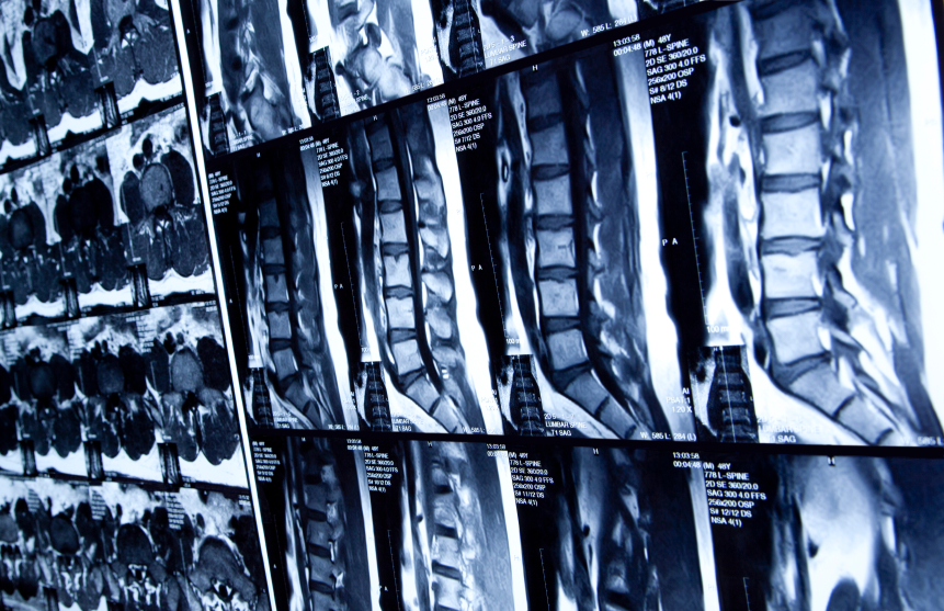
MRI technique detects spinal cord changes in MS patients: study
A Vanderbilt University Medical Center-led research team has shown that magnetic resonance imaging (MRI) can detect changes in resting-state spinal cord function in patients with multiple sclerosis (MS). Read MoreApr 19, 2018
-
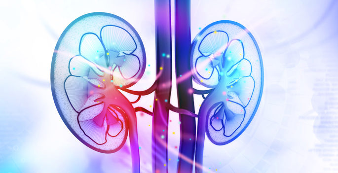
Kidney disease imaging
Making multiple measurements with MRI can provide comprehensive information about the molecular and cellular changes caused by kidney injury. Read MoreMar 22, 2018
-
New imaging approach offers unprecedented views of staph infection
A new integrated imaging approach makes it possible to probe the molecules involved in invasive infections and can be broadly applied to any health or disease state. Read MoreMar 15, 2018
-
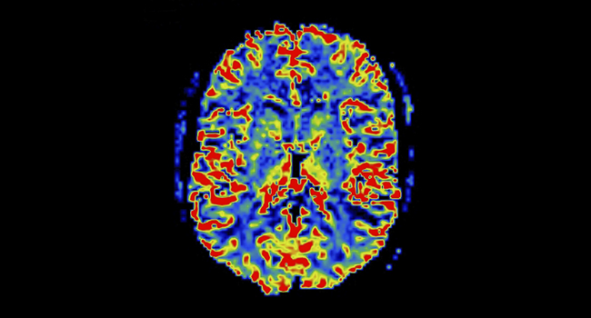
BOLD view of white matter
Vanderbilt investigators have discovered that functional MRI detects neural activity in both gray and white matter in the brain, suggesting new ways to investigate diseases such as Alzheimer’s and multiple sclerosis. Read MoreJan 12, 2018
-
Study finds common brain scanning technique maps electrical activity as precisely as more invasive methods
A commonly used brain scanning technique can map electrical activity under the skull as precisely as more invasive methods that rely on probes or electrodes, researchers at Vanderbilt University Medical Center (VUMC) reported this month. Read MoreMay 25, 2017
-

Pettigrew lecture
Roderic Pettigrew, Ph.D., M.D., right, director of the National Institute of Biomedical Imaging and Bioengineering of the National Institutes of Health, poses for a photo with Vanderbilt’s John Gore, Ph.D., left, and André Churchwell, M.D., following his recent Flexner Discovery Lecture. Read MoreMar 9, 2017
-
Study reveals biomarker of post-injury spinal cord function
Vanderbilt University researchers have demonstrated, for the first time in a primate model, that injury disrupts neural signaling in the spinal cord and that these changes can be measured non-invasively with functional magnetic resonance imaging (fMRI). Read MoreApr 23, 2015
-

‘White matter’ behaves differently in children with dyslexia
Trans-institutional neuroimaging research at Vanderbilt finds that the brain may be structured differently in children with dyslexia. Read MoreOct 29, 2014
-

Technique brings spinal cord neural signaling into focus
Researchers in the Vanderbilt University Institute of Imaging Science have achieved the first conclusive non-invasive measurement of neural signaling in the spinal cords of healthy human volunteers. Read MoreAug 5, 2014
-

Gore honored by Zhejiang University
John Gore, Ph.D., director of the Vanderbilt University Institute for Imaging Science, was named an honorary professor of Zhejiang University, China, during his recent visit to Zhejiang University School of Biomedical Engineering and Instrument Science. Read MoreFeb 13, 2014
-
Academy of Radiology Research honors VU’s Gore, Omary
Two leaders in imaging science at Vanderbilt University are among 43 recipients of the 2013 Distinguished Investigator Award from the Academy of Radiology Research, academy officials announced last week. Read MoreJun 27, 2013
-
Team creates new view of body’s infection response
A new 3-D view of the body’s response to infection — and the ability to identify proteins involved in the response — could point to novel biomarkers and therapeutic agents for infectious diseases. Read MoreAug 9, 2012
-

Minds wide open: Neuroscience at Vanderbilt
Vanderbilt University has emerged as one of the nation’s leading academic centers in neuroscience. Read MoreApr 6, 2012