Reading is a gateway skill.
Studies show that children who have serious difficulty and fail to master it are more likely to drop out of high school, lack meaningful employment and wind up living in poverty.
That is what makes Laurie Cutting’s research so vital. For 15 years, the Patricia and Rodes Hart Professor of Special Education, and professor of psychology and human development, radiology and pediatrics, has been working to identify and characterize the brain’s reading network—specifically how it may be different in those who have difficulty learning to read as compared to typically developing readers.
The numbers vary depending on how it is defined, but Cutting estimates that around 10 percent of the population suffer from a reading difficulty of some kind. Of those, about half can improve their reading skill with scientifically based remedial instruction. For the other half—while it is not understood exactly why they don’t respond to intervention—their disability could be neurobiological.
“We want to understand the different systems in the brain that support reading so we can identify the causes of different types of reading disabilities and then use that knowledge to improve remediation efforts,” Cutting said.
In pursuit of this goal, she has combined different disciplines that until recently have had little overlap: the field of education and the highly technical field of brain imaging.
“To my mind, Laurie is one of a handful of people in the world who have such a depth and breadth of knowledge in two domains: the world of cutting-edge neuroscience and the arena of educational practices and procedures,” said Mark Wallace, director of the Vanderbilt Brain Institute and professor of hearing and speech sciences.
This interdisciplinary ability has allowed her to become a major conduit of collaboration between the educational specialists at Peabody College and the biomedical engineers at the Vanderbilt University Institute of Imaging Science—one of the top centers for magnetic resonance imaging research in the country.

The Evolution of Magnetic Resonance Imaging
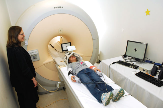
When Cutting began her career in the mid-1990s, MRI was coming of age as an exciting new research tool. For the first time it provided neuroscientists with a safe and unobtrusive way to understand brain structure and function in living people. MRI uses strong magnetic fields and radio waves to form images of the soft tissue in the body. At first it provided new information on basic brain structure.
Then engineers developed a variant called functional MRI, or fMRI, which provides information on the transient changes in blood-oxygen levels in different regions of the brain, making it possible to map changes in brain activity.
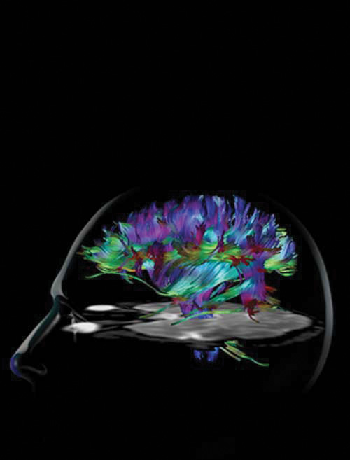
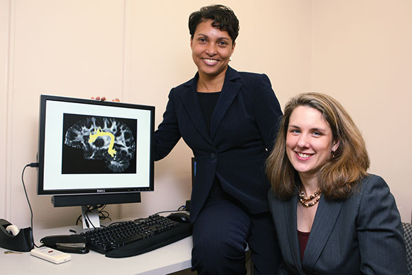
Enter diffusion MRI, yet another adaptation that measures the movement of water molecules in biological tissues. Neuroscientists use the technique to study how the different parts of the brain are wired together. It allows them to trace the paths and estimate the density of the bundles of nerve fibers that make up the “white matter” in the brain.
Early on, Cutting recognized the value of this technology for studying reading disabilities and received a postdoctoral fellowship at Johns Hopkins Medical School to learn how to apply it.
“Laurie’s become very experienced at imaging the developing brain,” said John Gore, director of VUIIS and University Professor of Radiology and Radiological Sciences. “She has had a number of unique ideas and done original work that has advanced understanding of the brain’s reading circuits and how to measure
the effects that different interventions have
on them.”
Mapping the Brain
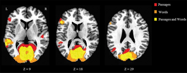
Adam Anderson, associate professor of biomedical engineering and an MRI expert at VUIIS, conducts brain mapping with Cutting. They have used fMRI to examine the reading network’s widely distributed nodes in the outer layer of the brain (the cortex), which handle different parts of the process. The reading network is a subset of the language network and it consists of four primary areas in the
left hemisphere:

- an area which recognizes the visual forms the occipital lobe;
- an area where letters are associated with sounds, in the supramarginal gyrus on the parietal lobe;
- the speech production center, in the inferior frontal gyrus on the frontal lobe; and
- an area where meaning is attached to words, in the angular gyrus on the parietal lobe.
The four regions are heavily interconnected with nerve fiber bundles. They are also connected to areas deeper in the brain including the thalamus, which regulates consciousness, alertness and sleep. In addition, higher-level reading processes invoke a much larger network of regions that are not language-specific and span both hemispheres.
“The brain didn’t evolve to be a reading machine,” Anderson said. “Reading is a skill that we have acquired quite recently. So it is piggy-backed on much older systems that evolved to do different things. That may be why reading disabilities are so prevalent.”
Not all Reading Disorders are Alike
Assistant Professor of Pediatrics Sheryl Rimrodt, who is a specialist in neuro-developmental disabilities, is another one of Cutting’s long-time collaborators. “I spend a lot of time in my clinics reassuring patients that their brains are not broken, their brains just work differently,” Rimrodt said.
Neuroimaging, she pointed out, has reinforced the view that not all reading disabilities are the same. They come in many different flavors. This means that reading instruction must be individualized. Instructors must identify the specific areas where a child is having difficulty and tailor the instruction to repair these gaps.
For example, children with dyslexia confuse letters and struggle to sound out words. Cutting’s research has shown that the connections between the area of the brain that specializes in visual word formation and the other linguistic centers are weaker in children with dyslexia than in typically developing readers.

Specific reading comprehension deficit, or S-RCD, is another common but less-known reading disorder.
Unlike children with dyslexia, children with S-RCD can read fluently but have difficulty comprehending what they are reading.
Cutting’s fMRI studies of children with this disorder have shown typical activity levels in the visual word form area but deficits in regions associated with memory.
“It may be that these individuals have a whole different neurobiological signature associated with how they read that is not efficient for supporting comprehension,” Cutting said.
Yet another type of reading disability is called late emerging reading disability, or LERD. In this case, children have no trouble reading until about the fourth grade, when they begin having trouble with both word formation and comprehension.

“School teachers have known about this for some time. They called it the ‘fourth grade slump,’ but it is more than that,” said Rimrodt. LERD accounts for between one-fifth to one-half of reading disorders identified late in elementary school.
Cutting wants to find a distinctive neurobiological signature for this type of disorder.
LERD occurs at the time when children are moving from reading stories to reading for comprehension, along with certain development in prefrontal cortices. It may involve deficits in what the researchers call the “executive function,” a collection of cognitive skills such as memory, attention control, organizational skills and a number of other functions not specific to reading, which are thought to be subserved by prefrontal cortices.
“The involvement of the executive function in reading is relatively uncharted territory,” said Cutting. “We have made some pioneering contributions by determining that it may have a relationship with an individual’s ability to comprehend what they are reading, and perhaps to compensate for shortcomings and to respond to intervention—but there’s a great deal more to learn.”
Finding the Right Intervention

Another focus of Cutting’s research is finding neurobiological signatures that predict how well individuals with reading disabilities will respond to remedial training. Laura Barquero, one of several doctoral students currently working and studying in Cutting’s Education and Brain Sciences Research Lab, is pursuing this as her thesis.

“We have found differences in the brain scans of those who respond to intervention and those who don’t,” Barquero said. The study took 23 children with reading disabilities, put them through an fMRI scanner, gave them 15 hours of reading intervention and tested the effect it had on their reading level. Barquero and her colleagues then compared the brain scans with the test results and found they could distinguish between the two groups.
Through the efforts of Cutting and her peers, interdisciplinary studies of reading development have become the leading wedge for efforts to address the broader question of the role that educational experiences play in shaping the specific brain circuitry that produces complex skills such as reading, writing and arithmetic. So it is only natural that Cutting has become a key player in one of Vanderbilt’s newest educational initiatives.
Cutting, with the Brain Institute’s Mark Wallace and Assistant Professor of Psychology Gavin Price, established the nation’s first educational neuroscience doctoral program in 2012. A partnership between Peabody and the Vanderbilt Brain Institute, the program furthers scientific understanding in the areas of child development, educational assessment, educational intervention and family processes.
The program also partners with the Vanderbilt Kennedy Center, the Center for Integrative Neuroscience and the Institute of Imaging Sciences. Key faculty from the departments of special education, hearing and speech sciences and psychology and human development are collaborators in this unique trans-institutional partnership.
“Vanderbilt has tremendous strengths in both neuroscience and education,” Wallace said. “It’s a natural progression to merge these strengths to build a world-class program.”
Learn more at vu.edu/cuttinglab or vu.edu/edneuro.