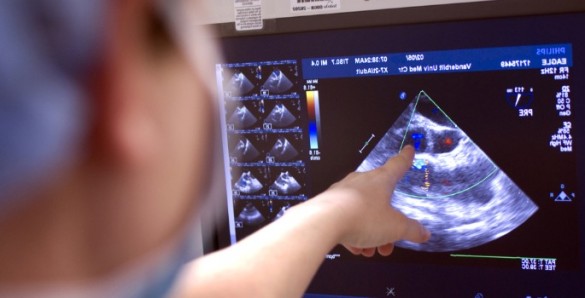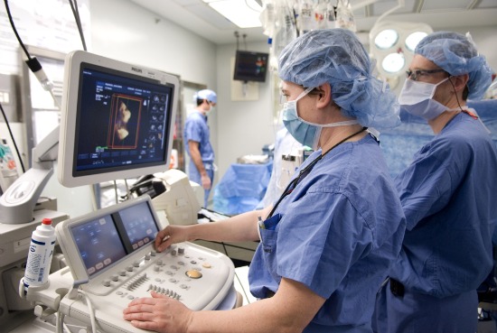
Vanderbilt cardiothoracic anesthesiologists and surgeons are pioneering the use of a tool that many in the cardiac field are calling the “new stethoscope” when it comes to monitoring critically ill patients.
Transesophageal echocardiography (TEE) is a diagnostic procedure that involves feeding an ultrasound probe through a patient’s mouth and into their esophagus to evaluate heart function. Because the esophagus is close to the heart, TEE can generate high-resolution images as the organ pumps. TEE is invaluable in locating cardiac blood clots, masses and tumors and can detect the severity of valve problems, congenital heart diseases and aortic tears.
Vanderbilt’s cardiothoracic anesthesiologists have more than 15 years of experience using TEE for patients undergoing cardiac surgery. Now, smaller, disposable TEE probes are being used to monitor the hearts of critically ill patients in intensive care units for as long as three days.
“The ability to monitor cardiac function and filling in a serial fashion with miniaturized, disposable TEE probes leads to optimal patient outcome and appropriate resource utilization,” said Chad Wagner, a cardiothoracic anesthesiologist and director of the Cardiovascular Intensive Care Unit. “The only real barrier to the routine use of this technology is physician training.”
‘Flying with instruments’
Using TEE to monitor cardiac patients in ICUs both before and after surgery allows medical staff to quickly intervene in a crisis and use the resulting information to guide treatment decisions.
“This technology is another example of what we describe as ‘flying with instruments,’ that is, the systematic approach of measuring and recording outcomes so as to guide further care,” said John G. Byrne, chairman of the Department of Cardiac Surgery.

To train physicians in the use of TEE, Wagner and Julian Bick, a cardiothoracic anesthesiologist, recently directed a course at Vanderbilt’s Center for Experiential Learning & Assessment that attracted physicians from premier health care systems across the United States, including two critical care doctors from the U.S. Navy.
Participants trained using a TEE simulator, which combines a photo-realistic, three-dimensional computer-generated model, a TEE probe and a simulated ultrasound image to help users visualize and understand complex cardiac anatomy. Bick also received a $100,000 grant from the Foundation for Anesthesia Education and Research to fund TEE simulation training for Vanderbilt University School of Medicine anesthesia resident physicians.
“The TEE simulator is highly effective in helping physicians learn TEE in combination with clinical training,” Bick said. “The simulator mirrors cardiac anatomy and function, and students often comment on the realism.”
Bick and Wagner, in collaboration with Byrne, recently published the first case study on using a disposable TEE probe to care for a 66-year-old patient who became unstable in the ICU following open heart surgery.
“Sometimes, unstable patients need to return to the operating room for additional surgery,” said Wagner. “In this case, after placing the miniaturized disposable TEE probe I was able to diagnose and treat the cause of the patient’s low blood pressure, which did not require returning to the operating room.”
Efforts to train more physicians to use TEE to care for the critically ill are already under way.