Microscopy
-

Vanderbilt researchers find evidence that the hunger hormone leptin can direct neural development in a leptin receptor–independent manner
Researchers from the Vanderbilt University School of Medicine Basic Sciences have uncovered the first example of activity-dependent development of hypothalamic neural circuitry. Read MoreDec 5, 2024
-

Protein dynamics in the beating heart
To study the dynamics of structural proteins in the heart, Vanderbilt investigators generated a cellular tool they expect will be useful for screening drugs that affect heart muscle contraction. Read MoreDec 16, 2019
-
Advanced imaging tools reveal architecture of cell division machinery
Using super-resolution microscopy tools in the Nikon Center of Excellence, Vanderbilt investigators have determined the molecular architecture of the contractile ring machinery that functions during cell division — a process that is essential for life. Read MoreNov 9, 2017
-

Nikon Center of Excellence makes debut Oct. 4
On Tuesday, Oct. 4, the Cell Imaging Shared Resource (CISR) at Vanderbilt University Medical Center (VUMC) will officially unveil its new Nikon Center of Excellence, which will feature state-of-the-art microscopy for live cell imaging. Read MoreSep 29, 2016
-
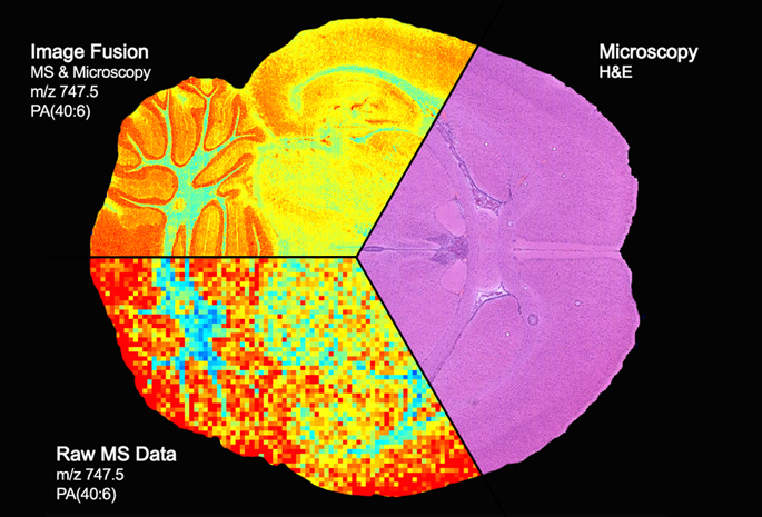
Vanderbilt team first to blend high-end imaging techniques
Vanderbilt University researchers have achieved the first “image fusion” of mass spectrometry and microscopy — a technical tour de force that could, among other things, dramatically improve the diagnosis and treatment of cancer. Read MoreMar 5, 2015
-

Pioneers of Discovery: Investigator taps into artistic side to reveal cells’ secrets
Dylan Burnette, Ph.D., points to one of the many striking photographs on his office walls. It’s a picture of a cell — a microscopic image showing yellow squiggles, bright purple lines and a turquoise oval on a black background, and it looks like abstract art. Read MoreMay 29, 2014