Cell Imaging Shared Resource
-
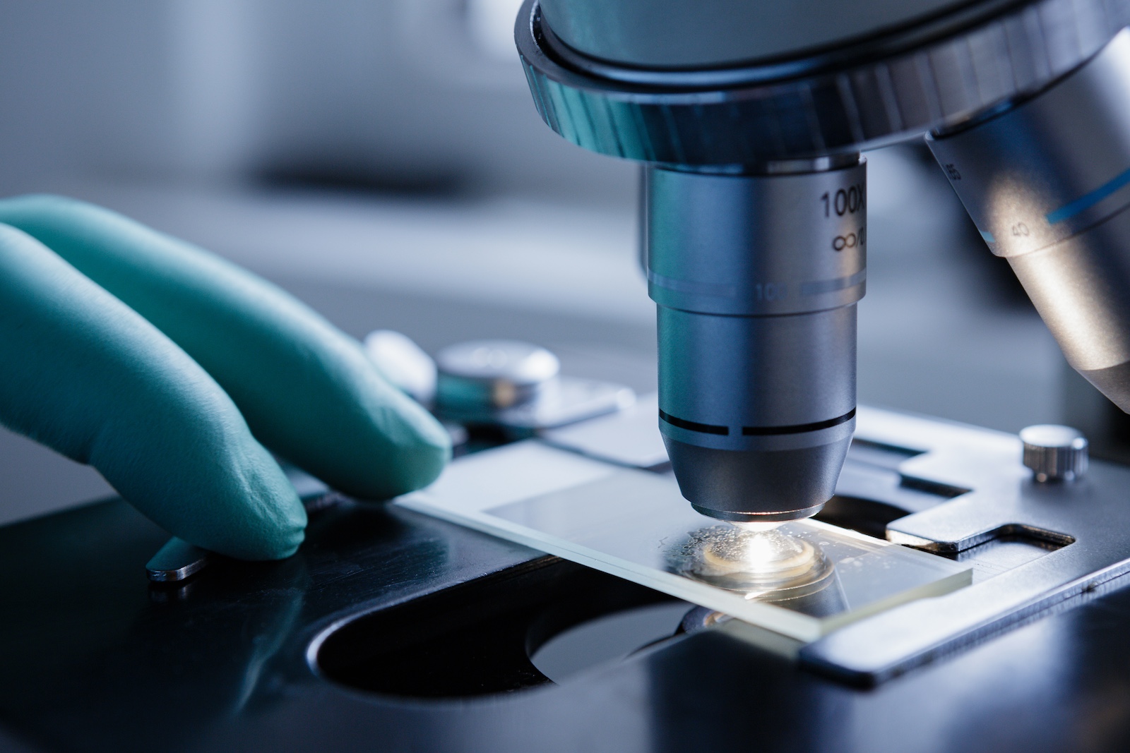
Aging researchers find new puzzle piece in the game of longevity
Think of cells as factories that hold sets of machines doing different things. How those machines are organized and used determines the efficiency of the factory. Vanderbilt researchers are looking into how cells reorganize those machines over time—and what that means for aging. They’re focused on a cell structure (machine) called the ER, which is known to be vital to cell processes but has not yet been thoroughly studied. “Changes in the ER occur relatively early in the aging process,” says Assistant Professor Kris Burkewitz. “One of the most exciting implications of this is that it may be one of the triggers for what comes later: dysfunction and disease.” And identifying the trigger could lead to being able to stop the firing. Read MoreMar 12, 2026
-
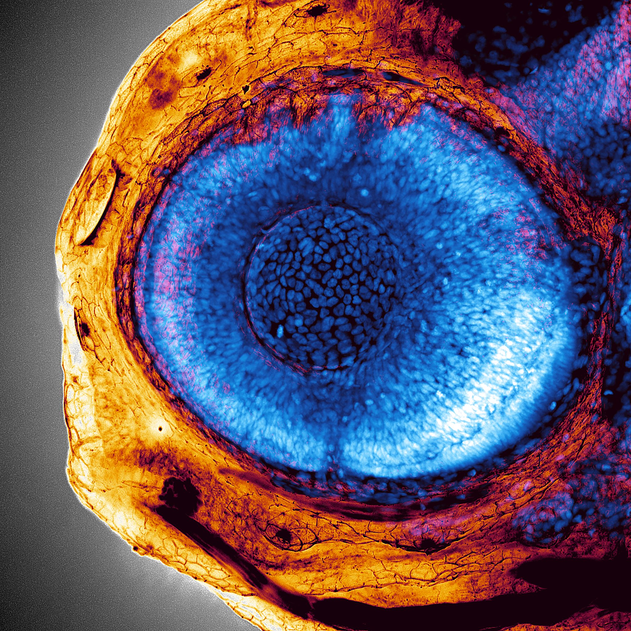
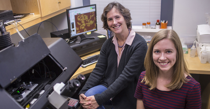
Hayes, Nagarajan and Costanzo win 2025 Cell Imaging Shared Resource Life Is Beautiful Image Contest
James Hayes, Rekha Nagarajan and James Costanzo are the winners of the 2025 Cell Imaging Shared Resource Life Is Beautiful Image Contest. They worked with Jenny Schafer, CISR managing director and research associate professor of cell and developmental biology, Oleg Kovtun, research assistant professor of chemistry, and CISR senior research specialists Kari Seedle and Tegy Vadakkan to create these images as part of their individual research projects. Read MoreAug 13, 2025
-

Protein dynamics in the beating heart
To study the dynamics of structural proteins in the heart, Vanderbilt investigators generated a cellular tool they expect will be useful for screening drugs that affect heart muscle contraction. Read MoreDec 16, 2019
-
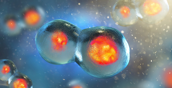
Understanding cell division
Vanderbilt researchers have uncovered another piece in the puzzle of how cells divide — a process that goes awry in cancer cells. Read MoreNov 18, 2019
-
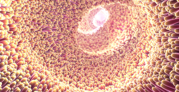
Microvilli in motion
Live cell imaging studies have revealed that microvilli — finger-like protrusions on the surface of epithelial cells — move and collide as they form the brush border. Read MoreSep 19, 2019
-
Advanced imaging tools reveal architecture of cell division machinery
Using super-resolution microscopy tools in the Nikon Center of Excellence, Vanderbilt investigators have determined the molecular architecture of the contractile ring machinery that functions during cell division — a process that is essential for life. Read MoreNov 9, 2017
-

Discovery sheds new light on Angelman, Prader-Willi syndromes
A mutation associated with epilepsy and autism also is responsible for a “pale eye” trait in two rare genetic disorders, Angelman syndrome and Prader-Willi syndrome, neuroscientists at Vanderbilt University Medical Center reported this week. Read MoreDec 22, 2016
-
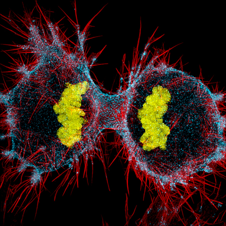
Striking view
Dylan Burnette, Ph.D., assistant professor of Cell and Developmental Biology, won 12th Place in Nikon’s Small World 2016 Photomicrography Competition for a colorful image of a dividing cancer cell. Read MoreOct 27, 2016
-
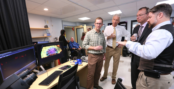
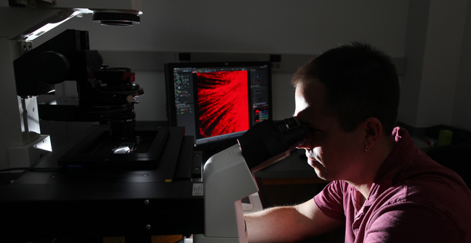
Nikon Center of Excellence for live-cell imaging makes debut
Officials of Vanderbilt University, Vanderbilt University Medical Center (VUMC) and Nikon Instruments Inc. last week celebrated the opening of the Vanderbilt Nikon Center of Excellence, which features state-of-the-art microscopy for live-cell imaging. Read MoreOct 13, 2016
-
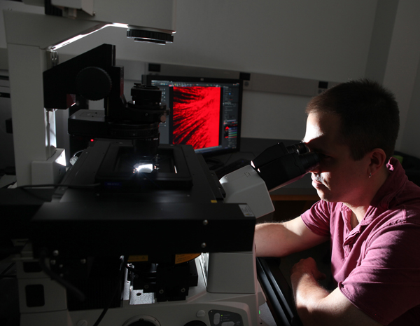
Nikon Center of Excellence makes debut Oct. 4
On Tuesday, Oct. 4, the Cell Imaging Shared Resource (CISR) at Vanderbilt University Medical Center (VUMC) will officially unveil its new Nikon Center of Excellence, which will feature state-of-the-art microscopy for live cell imaging. Read MoreSep 29, 2016
-
VUMC research cores speed pace of discovery
Progress against America’s most intractable health challenges, among them heart disease, cancer and diabetes, requires the best minds, the latest tools and the easy collaboration demanded by 21st century science. Read MoreJan 22, 2015
-
New ‘super’ microscopes sharpen cellular imaging
Two new “super-resolution” optical microscopes have put Vanderbilt University Medical Center on the cutting edge of cellular imaging, and are giving researchers their first views of the cell at the molecular level. Read MoreJul 11, 2013