


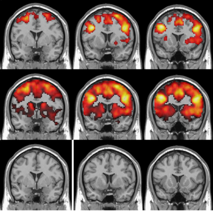
Brain activity requires higher rates of oxygen delivery and blood flow, which increase BOLD (blood oxygenation-level dependent) signals that are detectable by functional magnetic resonance imaging (fMRI).
BOLD signals have usually been associated with activity in the gray matter of the brain — where neuronal cell bodies reside. Growing numbers of studies have detected BOLD signals in white matter, the bundles of nerve fibers (axons) that transmit signals, but no comprehensive measurements have been made to characterize the signals.
Muwei Li, PhD, and colleagues in the Vanderbilt University Institute of Imaging Science, have used an event-related cognitive task (Stroop color-word interference) to measure the BOLD hemodynamic response function in specific human white matter pathways. Reporting in Nature Communications, they showed reduced magnitudes and delayed responses in white matter compared with gray matter.
The findings demonstrate the detectability of neural activities in white matter and point to the need for changes to models for assessing white matter activity and physiology.
This research was supported by National Institutes of Health grant NS093669.