


Elizabeth and Scott Massey expected to learn their baby’s gender at a 21-week ultrasound. While they learned they were having a daughter, the reveal included much more than they planned for.
Multiple ultrasounds and a fetal MRI showed their baby, whom they named Charlotte, had a host of internal physical irregularities: a partially formed spine, with a missing section of the thoracic vertebrae; she only had three of the 12 ribs that protect and support the left lung, which consequently never developed to full size; her heart was incorrectly positioned on the right side of her chest; she had one central kidney and an intestinal obstruction. She also had a large skin-covered meningocele, where the membranes that cover the spine and part of the spinal cord protrude through a defect in the vertebral column.
For the Masseys, from Milan, Tennessee, the radiology images taken while Elizabeth was pregnant were hard to process and fully comprehend.
As an educational tool for the Masseys, as well as to inform care decisions among her team of doctors, the Vanderbilt Department of Radiology and Radiological Sciences’ 3-D Printing Center, led by Sumit Pruthi, MD, created a 3-D model of Charlotte’s internal anatomy, specifically highlighting the spine, ribs and lungs.
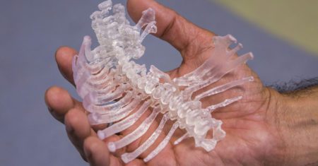
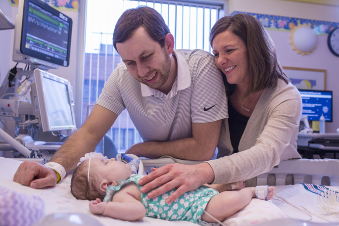
“When we had a care conference with the model we were able to get a better understanding and an actual visual of what the ribs look like, especially on the right side where she has them and where they are absent on the left side,” Scott Massey said. “We could hold it and see that this is what our daughter’s anatomy looks like.”
Elizabeth Massey added, “To hear her ribs are fused together: well what does that mean? (The model) helps to give a clearer picture of her story. We were able to ask questions we might not have considered.”
The recently launched 3-D Printing Center is located on the first floor of Monroe Carell Jr. Children’s Hospital at Vanderbilt and serves both pediatric and adult cases.
Pruthi helped establish the center with the support of Children’s Hospital administration and of Reed Omary, MD, chair of Radiology.
The effort is also supported by Allen Newton, PhD, from the Vanderbilt Institute of Imaging Sciences, Hansen Bow, MD, from Neurosurgery, Jason Christensen, MD, from Pediatric Cardiology, and Steven Lewis, lead engineer of the program.
The 3-D Printing Center currently has three 3-D printers that have the ability to create prints, or three dimensional models, ranging in size from the width of a human hair to the length of a leg. A structured light-based 3-D scanner is also part of the equipment, and they use Materialise Mimics Innovation Suite, which is currently the only FDA medically cleared 3-D printing software. The center has produced more than 56 models since January, catering to different departments and different needs.
“One of the goals at Vanderbilt is innovation, and this technology certainly is in keeping with that mission. 3-D printing is innovative and part of the future of medicine,” said Pruthi, associate professor of Radiology and Pediatrics and chief of Pediatric Neuroradiology. “The models can be used for patient education, medical student and resident education/training, surgical planning and simulation. If a person has a tumor with complex surgical anatomy, we can make a 3-D tumor model which the surgeon can hold in his/her hand, in order to optimize surgical planning. Models can also be used to practice complex steps in operations before they are performed.”
Pruthi said anyone at Vanderbilt can request 3-D models via a link on eStar. He, along with Steven Lewis, who sees each model through to completion, emphasizes the importance of discussing the goals of the project and determining anatomical regions that need to be segmented in order to refine a model’s purpose and maximize its utility. Currently, models can be created from one of about four different medical and dental materials with a range of colors and textures, a number which Pruthi hopes to expand.
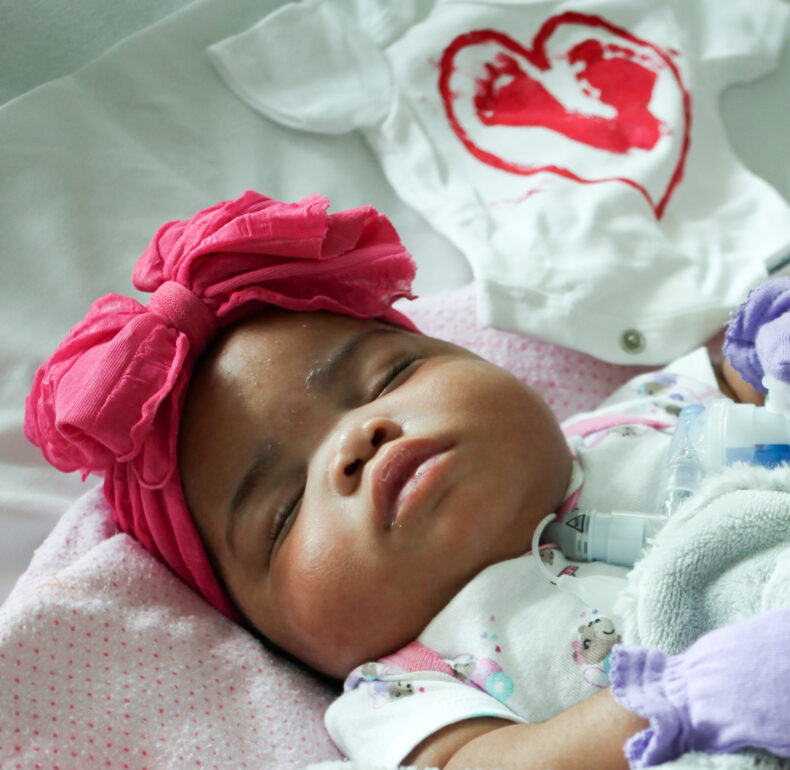
In Charlotte’s case, her healthcare team wanted to fully understand the best way to care for her after birth, since her chance for survival was considered limited. Born Dec. 8 via cesarean section, Charlotte let out two loud cries before she was whisked off to the NICU where she was intubated to help her breathe.
Four days later, Jay Wellons, MD, chief of Pediatric Neurosurgery, successfully repaired Charlotte’s meningocele from her back, which was larger than her newborn head. Soon after, her pediatric surgeon, Harold Lovvorn III, MD, requested the model of her internal anatomy in time for a team care conference with the parents. Lovvorn performed two abdominal procedures on Charlotte.
“It really is helpful to hold the model in one’s hand to look at what the anatomy and malformations are before actually operating on a patient,” said Lovvorn, associate professor of Pediatrics and Pediatric Surgery. “In Charlotte’s case, it was also an excellent teaching tool for the family — and really for everybody on her care team. The model solidified our thinking.”
The team explored if there was an option to give her prosthetic ribs — unlikely due to lack of a portion of vertebral column to which they could attach.
The model also allowed the caregivers to discuss options for correcting Charlotte’s severe scoliosis as she grows.
“The more information we have as care providers and the more precise that information is, not only can we perform better operations and provide better care, but we can also counsel a family better and they can have a better appreciation of what their baby or child is up against,” Lovvorn said.
Charlotte, now almost 4 months old, will go home when she gets a little bigger and she will be transitioned to a home ventilator for breathing. The Masseys also have a copy of the 3-D model which they can show to other caregivers who will take care of Charlotte in the future.