


The vast majority of meningiomas — tumors that form from the membranes surrounding the brain and spinal cord — are slow-growing and benign. “Atypical” meningiomas have a more aggressive clinical course, and patients with atypical tumors would potentially benefit from earlier surgery and efforts to achieve complete tumor removal.
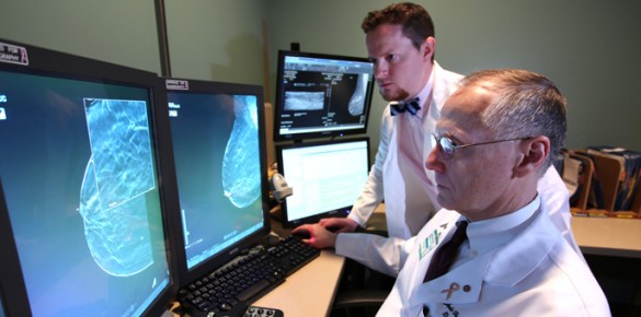
Andrew Hale, an MD/PhD candidate at Vanderbilt, Lola Chambless, MD, and colleagues reviewed magnetic resonance imaging (MRI) studies for 128 patients who had benign or atypical meningiomas surgically removed.
They found that tumor volume was the most striking single predictor of tumor grade. Additional imaging features associated with increased risk for atypical pathology included the presence of tumor necrosis, swelling around the tumor (peritumoral edema) and tumor location.
The findings, reported in the February issue of the Journal of Clinical Neuroscience, demonstrate the contribution of tumor volume to atypical meningioma pathology and may help guide surgical planning and patient counseling.
Hale is supported by the MD/PhD training grant funded by the National Institutes of Health (GM007347).
Send suggestions for articles to highlight in Aliquots and any other feedback about the column to aliquots@vanderbilt.edu