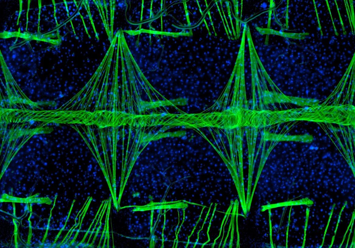
A fluorescent image of the heart of a mosquito taken by a Vanderbilt graduate student has captured first place in Nikon’s “Small World” 2010 photomicrography competition.
Jonas King took the image that shows a section of the tube-like mosquito heart magnified 100 times. He is a member of the research group of Julián Hillyer, assistant professor of biological sciences, and the image was taken as part of their research on the circulatory system of Anopheles gambiae, a mosquito that spreads malaria.
According to Nikon, 2,200 images were submitted—the largest number in the 36-year history of the competition—and King’s image was judged the winner for its combination of aesthetic beauty, scientific relevance and the technical difficulty involved in capturing it.
“[[rquote]Surprisingly little is known about the mosquito’s circulatory system despite the key role that it plays in spreading the malaria parasite,” Hillyer said.[/rquote] “Because of the importance of this system, we expect better understanding of its biology will contribute to the development of novel pest- and disease-control strategies.”
The mosquito’s heart and circulatory system is dramatically different from that of mammals and humans. A long tube extends from the insect’s head to tail and is hung just under the cuticle shell that forms the mosquito’s back. The heart makes up the rear two-thirds of the tube and consists of a series of valves within the tube and helical coils of muscle that surround the tube. These muscles cause the tube to expand and contract, producing a worm-like peristaltic pumping action.
Most of the time, the heart pumps the mosquito’s blood—a clear liquid called hemolymph—toward the mosquito’s head, but occasionally it reverses direction. The mosquito doesn’t have arteries and veins like mammals. Instead, the blood flows from the heart into the abdominal cavity and eventually cycles back through the heart. “The mosquito’s heart works something like the pump in a garden fountain,” Hillyer said.
To show the structure of the mosquito heart, King used two types of fluorescent dye. The green dye binds with muscle cells and shows the underlying musculature. The blue dye binds with cellular DNA and shows the presence of all the mosquito’s cells. The point of view of the image is top down. The mosquito’s body lies horizontally with its head to the left. The heart is the narrow tube that runs horizontally across the middle of the picture. The muscles that wind around the heart show up clearly in green. The triangular-shaped bundles perpendicular to the heart are called alary muscles and they hold the heart up against the mosquito’s back. Each of these bundles is centered on one of the heart valves, which do not show up clearly. The mosquito’s body consists of a series of segments and the broad strips of muscle that run parallel to the heart are intersegmental muscles that hold the segments together. The vertical muscles at the top and bottom of the image wrap around the mosquito’s body and are called intrasegmental muscles.
More information and additional images are available on the Hillyer Lab website. Hillyer’s research is funded by grants from the National Science Foundation.