


by Sanjay Mishra
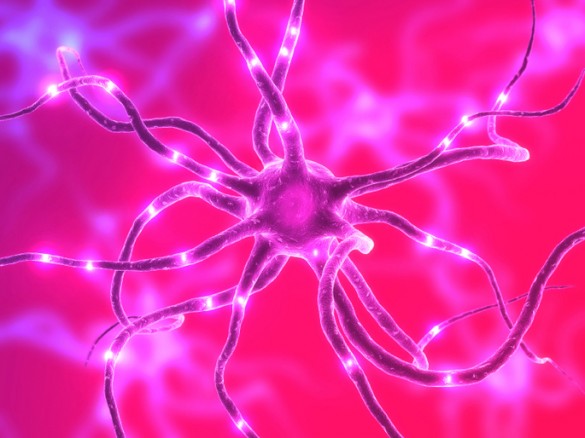
The brain is comprised of gray and white matter, made of neurons with axon projections.
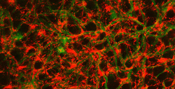
In mainly white matter, myelin wraps to form a sheath around axon fibers. This accelerates nerve signals by electrically insulating the axon. Many white matter diseases involve loss of myelin, which makes diagnosing the health of myelin clinically very important.

The ratio of the axon diameter to diameter of the axon and myelin sheath, called g-ratio, can be used as a good measure of the overall health of white matter. However, current methods of assessing g-ratio are time-consuming and invasive.
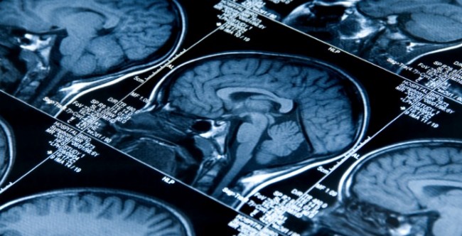
Magnetic resonance imaging (MRI) is a promising alternative, but the current models of estimating g-ratio from MRI rely on the incorrect assumption that the g-ratio is constant for all axon fibers.
Reporting this month in the journal NeuroImage, Mark Does, Ph.D., and colleagues demonstrate a revised model for quantitatively estimating g-ratio from MRI. Their report shows that the g-ratio obtained from MRI is the axon-area-weighted measure of g-ratio across all axons.
This research was supported by the National Institutes of Health (grant EB001744).
Send suggestions for articles to highlight in Aliquots and any other feedback about the column to aliquots@vanderbilt.edu