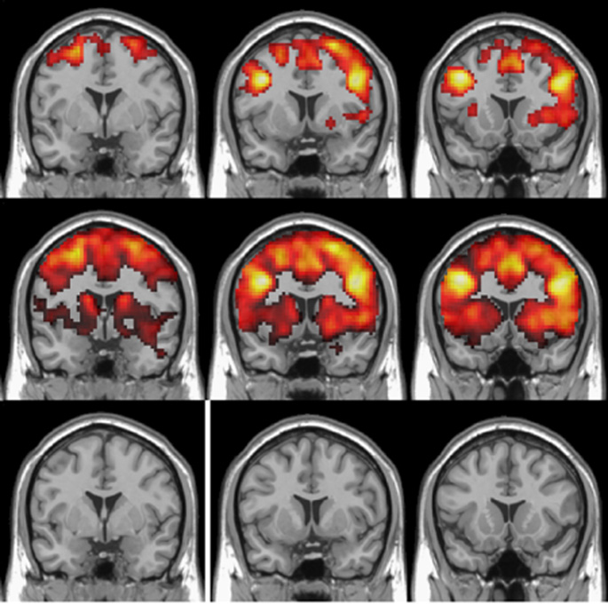


The hippocampus – a brain region involved in learning and memory – is abnormal in schizophrenia. Neuroimaging studies offer evidence of altered hippocampal activity, but mostly rely on a single hemodynamic parameter, for example blood volume or blood flow, and report mixed results.
Pratik Talati, a graduate student in Psychiatry, Swati Rane, Ph.D., research instructor at the Vanderbilt University Institute of Imaging Science, and colleagues used dynamic susceptibility contrast-MRI to simultaneously assess several hemodynamic measures – blood volume, blood flow and mean transit time (an indicator of circulation) – in the hippocampus of patients with chronic schizophrenia and healthy controls. They report in the June 30 issue of Psychiatry Research: Neuroimaging that patients with schizophrenia have significantly increased blood volume but normal blood flow and mean transit time in the hippocampus. The uncoupling of blood volume from flow could be due to antipsychotic medications or other factors, the authors suggest.
The findings support the use of multiple hemodynamic parameters to better link hippocampal function with clinical features of schizophrenia.
This study was supported by grants from the National Institutes of Health (MH070560, NS078828, MH102846, GM007347, RR024975).
Send suggestions for articles to highlight in Aliquots and any other feedback about the column to aliquots@vanderbilt.edu