Monroe Carell Jr. Children’s Hospital at Vanderbilt has become the first hospital in the state to install a new imaging system that will help to dramatically reduce X-ray exposure during certain heart procedures.
The imaging system allows doctors to rely on fluoroscopy images much less often during catheter ablation procedures used to correct heart arrhythmias for children and adults with congenital heart disease.
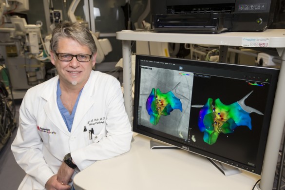
The new CartoUnivu system integrates the fluoroscopy images into a 3-D mapping program, allowing the physician to see in real time the location of the catheter during the procedure.
“This system allows you to utilize the anatomic landmarks you would normally need X-rays to use, particularly in patients with complex anatomy in whom the stand-alone Carto imaging is often inadequate,” said Frank Fish, M.D., professor of Pediatrics and director of the Pediatric Electrophysiology Laboratory at Children’s Hospital.
Until recently, fluoroscopy images were used heavily during heart ablation procedures to help the physician guide the catheter to the precise location inside the heart to perform the ablation, during which heat or cold is applied to the tissue to correct the arrhythmia.
Already, the new system has reduced the need for fluoroscopy by at least two-thirds on average, Fish said, thereby lowering radiation exposure for both the patient and the clinicians.
“Ultimately we hope to achieve fluoroscopy times for complex cases of 1 minute or less, compared to 15 to 30 minutes, which has been more typical for these cases,” Fish said. “While the radiation doses represented by these exposures are relatively low compared to other imaging exposures, such as CT scan or coronary angiography, there is a continued priority to reduce all exposures to ‘As Low As Reasonably Achievable’ (ALARA).”
The CartoUnivu system is manufactured by technology firm Biosense Webster and is part of the company’s Carto 3 electroanatomic mapping system, which is already in use at Children’s Hospital and Vanderbilt University Hospital for ablation procedures. The system incorporates other imaging modalities into the system as well, including cardiac MRI, CT angiograms, and intracardiac ultrasound.
“The installation of the CartoUnivu system is the final step in offering unsurpassed care to our young patients with arrhythmias,” said H. Scott Baldwin, M.D., Katrina Overall McDonald Professor of Pediatrics and chief of the Division of Pediatric Cardiology. “With Dr. Fish, we have one of the best Pediatric Electrophysiologists in the country, and he has assembled a superb team.”
The mapping system is one component of the hospital’s highly successful program to treat arrhythmias through catheter ablations. For the past six years, the program has had a 100 percent efficacy rate for catheter ablations to treat supraventricular tachycardia and a 96 percent success rate for more complex procedures in adult congenital heart disease patients. Overall, the procedures have reached a success rate of 99 percent.
“We can now provide this team with a tool that significantly decreases the potential risk from radiation incurred in long procedures thus making our EP lab not only one of best in terms of outcomes, but also one of the safest labs anywhere,” said Baldwin, also a professor of Cell and Developmental Biology.
While some medical centers have adopted systems that allow zero or minimal fluoroscopy for the procedures, Fish said those approaches have been less suitable for complex cases, in particular for adults with congenital heart disease.
“To date, we have been unwilling to compromise our procedural outcomes, but with this system, we can rely less on fluoroscopy on even our most complex cases,” Fish said.


